Kidney Stone

How is kidney stone formed?

Kidney stones are formed by the crystallization of various minerals such as calcium, oxalate and uric acid in the urine. Either the amount of minerals such as calcium, oxalate and uric acid increases, or the amount of substances such as citrate and magnesium that prevent stone formation in the urine decreases. The incidence of urolithiasis in Turkey is 11%, and Turkey is considered as an endemic area in the world.

Who are at risk for stone formation?

  • It is seen 2-3 times more in men. However, the gender gap between men and women has been closing in recent years. The incidence is equal in men and women between the ages of 1 – 18 and after 50.

Testesteron => increases oxalate synthesis and crystallization

Estrogen => increases citrate (prevents stone formation) excretion into urine


  • If there is a family history of stone disease, the risk is 2.5 times higher. Polygenic inheritance is in question, and some genetic disorders (RTA, cystinuria, etc.) may also cause stone formation.
  • Stone disease peaks between 4 and 6 decades.
  • Risk increases in hot and dry climate conditions (deserts, mountainous areas, tropical regions, etc.). In addition, stones are observed more frequently in those working in hot environments (due to dehydration) and in sedentary occupations
  • Obesity and sedentary life is also an important risk factor.

Is it related to food?

  • Yes, eating habits are associated with stone formation in some stone types. A balanced diet is essential.
  • Excessive protein consumption increases stone formation
  • Excessive salt consumption (sodium increase) paves the way for calcium crystallization in urine
  • Contrary to what is believed, calcium is not restricted from diet in patients with calcium-oxalate stones. Normal calcium consumption (1 – 1.2 g / day) is recommended. With calcium restriction, oxalate-Ca binding in the intestine decreases, the absorption of the released oxalate from the intestine increases, which increases the risk of stone formation.
  • Excessive consumption of oxalate-containing food and vitamin C (<2g / day) should be avoided (especially in those with hyperoxaluria)

What are the harms of kidney stones?

Apart from the fact that kidney stones can cause severe pain, they can cause kidney obstruction, permanent loss of kidney function and kidney failure.

By laying the groundwork for infection, it may cause dysfunction of other organs and cause severe infections (urosepsis).

What can be done to prevent stone formation?

Regular physical activity, adequate fluid consumption and a balanced diet are quite important. If you have urinary system stones, it would be useful to get information from your physician about your nutritional habits. It is important to investigate the underlying metabolic causes in patients with recurrent stone attacks, and to have regular check-ups in patients who require medical (drug) treatment.

How are kidney stones diagnosed?

While symptomatic stones cause pain and refer the patient to the physician, asymptomatic (silent) stones are often diagnosed incidentally.

We use biochemical and radiological methods to make a diagnosis. Urine analysis, blood tests are performed when necessary and the imaging method is decided. Ultrasonography is the primary diagnostic method and its most important advantage is that it does not contain radiation and is easy to apply. However, its sensitivity is low (45%). That means, it may not be able to detect the existing stone. Ultrasonography is the first-line diagnostic method in pregnant women and children.

The most sensitive imaging for stone detection is non-contrast computed tomography. With low-dose stone protocols, computed tomography can be performed with radiation as much as a lung x-ray.

What are the treatment options for kidney stones?

  • eSWL (shock wave lithotripsy): ESWL (stone breaking with shock wave): It is a treatment method that does not require anesthesia and is applied to small size kidney stones. This method is based on the principle of transmitting sound waves generated from a generator to the stone through the skin, thus breaking the stone. Several sessions may be required for the stone to be completely broken and poured. Broken stone fragments are spontaneously excreted from the body through the urinary tract.

It is not preferred in pregnant women, patients taking blood thinners, and patients with urinary tract infection, in the presence of obstruction in the urinary tract for some reason.

The hardness of the stone, the distance between the stone and the skin, the location and size of the stone in the kidney are factors affecting the success of the procedure.


  • F-URS(flexible-ureterorenoscopy): With thin devices (flexible ureteroscope) with a camera at the tip, the urethra and bladder are passed, the ureter is entered and the kidney is reached. Here, the stones are broken with the laser device and left to fall off by themselves.

Its biggest advantage is that it is performed without any incision in the body and the short hospital stay. In large-sized kidney stones, the possibility of complete purification from the stone is reduced.

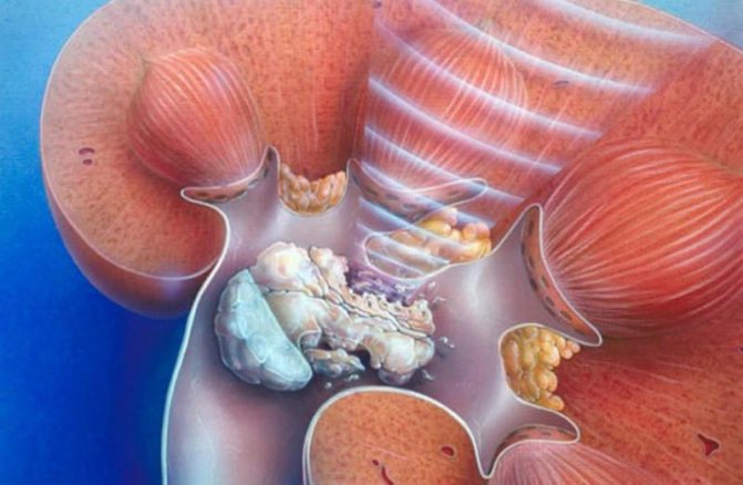
    • PNL (Percutan nephrolitotomy): This surgery is often performed under general anesthesia. During the operation, a path extending into the kidney from a 1cm incision from the back of the patients is formed and the camera is entered into the kidney with camera-assisted devices (nephroscope) through the tube placed on this path. Stones in the kidney are broken down and taken out.


    In case the stones fill different calyces of the kidney, it may not be possible to reach all stones from a single tube, despite the flexible devices you have. In such cases, it may be necessary to make more than one entry to the kidney. This additional intervention decision is a decision that the surgeon will make during the operation according to his experience. At the end of the procedure, a tube (nephrostomy) is mostly placed in the patient’s kidney to ensure a safe flow of urine and to resolve edema.

    The most important complication of the procedure is bleeding. Puncturing the kidney, which has a very dense blood supply, with a tube carries the risk of bleeding. But the structures around the kidney limit this bleeding in a short time. While blood transfusion is required in approximately 7% of patients after PNL operations; In 0.5%, bleeding that requires intervention occurs. This surgery is not performed when using the drug in patients with bleeding disorders or using anticoagulant or antiagregant drugs due to the risk of bleeding.

    One of the important complications of this surgery is infection. Therefore, in patients with urinary tract infections, this surgery should be delayed and should be done after the infection is completely cured.

    Despite all these, PNL surgery is one of the most reliable and effective treatment methods known today, especially for large stones.


    • Laparoscopic stone surgery: Laparoscopic method can also be applied safely in selected cases of kidney stones.


    What are the treatment options for ureteral stones?


    • Follow-up: 95% of stones up to 4 mm fall spontaneously within 40 days. Generally, follow-up up to 30 days is recommended (1st, 14th and 30th days are called for control). Follow-up is not recommended in case of infection, complete obstruction (worsening kidney function) and refractory pain. Addition of Medical Expulsive Therapy (alpha blocker drugs) is recommended because it will facilitate stone removal.
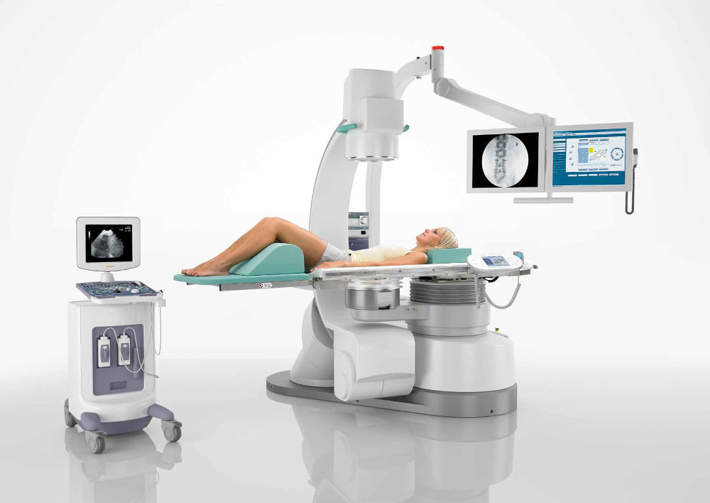
    • ESWL (stone breaking with shock wave): The effectiveness of ESWL in ureteral stones decreases as it descends from the kidney to the bladder. Again, as the size of the stone increases, the stone-free rates decrease.
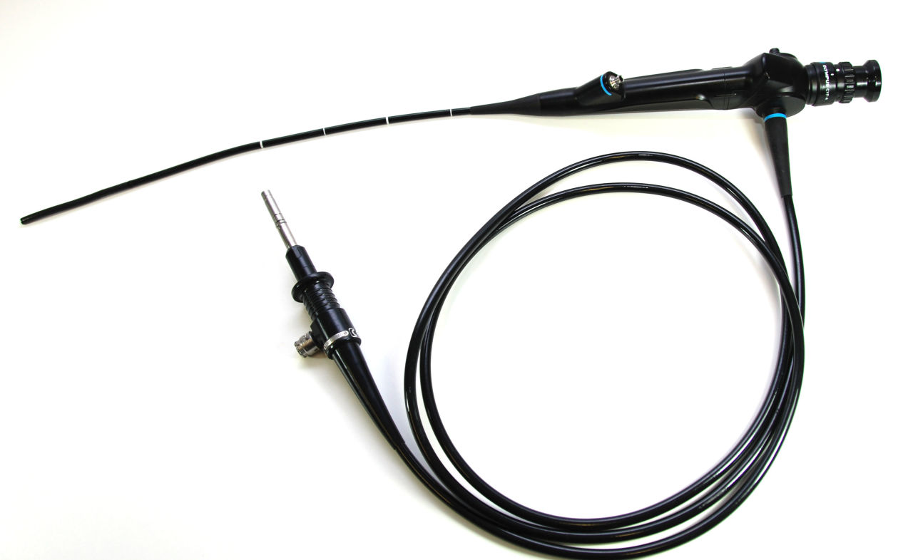
    • URS (ureterorenoscopy): Semirigitis or flexible ureteroscopes can be used in the treatment of ureteral stones. The success of URS increases as the stone size decreases and descends from the kidney to the bladder.
    • PNL (Percutaneous Nephrolithotomy): PNL has a limited place in ureteric stones. PNL is a method that can be successfully applied in proximal ureter stones.
    • Laparoscopic: The laparoscopic approach is a successfully applied method with high stone-free rates, especially in large ureteral stones.

    Treatment for kidney stones with different anatomy?

    In many congenital anomalies of the kidney such as horseshoe kidney, pelvic kidney, ectopic fusion kidney, kidney with rotation anomaly, the treatment of stone disease is special and experience is very important in the decision of treatment. In these patients, the most appropriate treatment option is applied according to the localization of the stone in the kidney, the localization of the kidney in the abdomen and the patient’s additional diseases.

    Which stones should be treated?

    If the ureter stones are large (stones larger than 1 cm are not expected to fall spontaneously, although there is no definite limit for a large definition), or if they completely obstruct the flow of urine, cause pain, have a single kidney or have stones in both ureters, surgical treatment is required for these stones. . In the presence of sepsis or pionephrosis (kidney filling with pus), after the infection treatment is completed by placing a stent (nephrostomy or ureteral stent), the stone surgery will significantly reduce the risk of the procedure.

    If the kidney stone is large (> 2 cm), causes pain, obstructs urine flow, and if the kidney is anatomically / functionally affected, these stones should be intervened. The localization of the stone in the kidney, the shape and location of the kidney, and additional disorders of the patient guide us to decide apropriate surgical method.

    Who should be investigated to detect the causes of stone formation?

    Metaboic evaluation should definitely be performed in childhood stone patients, patients with recurrent stone history, patients with single kidney stones, family history, and patients with suspected hereditary stone disease.

    Is there any drug treatment for stone disease?

    Drug treatments have a place in stone disease. Although there are treatments (medical expulsive therapy) available to facilitate the fall of ureter stones in this area, there are also treatment methods (medical prophylaxis) used to reduce the risk of stone recurrence. Preventive treatment can be planned according to the underlying metabolic cause and stone analysis result.