Prostate Cancer

What is cancer and how is it formed?

Cancer is the disruption and uncontrolled proliferation of cells that make up an organ or tissue. This uncontrolled reproduction distorts the shape of the organ and begins to grow over time. The rate of growth varies according to the structure of the organ or tissue and the type of cancer cells. Over time, they also spread to the surrounding tissues. If they enter the blood and lymph vessels, they also settle in other organs of the body, which is called metastasis.

Prostate cancer is the second most common cancer in men, and the number of patients diagnosed every year in the world is around 1 million. 15% of all prostate cancer cases are familial and hereditary. In these patients, cancer is usually detected at an early age (<55 years). Although prostate cancer susceptibility regions have been suggested on some chromosomes (often emphasized on chromosome 1), a precise gene localization has not been demonstrated. However, there are some articles demonstrated that there is a higher risk of prostate cancer for men with BRCA1/2 gene mutation in recent years.

cancer cell
onkolojik

Risk factors for prostate cancer

The most important risk factors for prostate cancer are: age, family history and race.

Prostate cancer is cancer of advanced age and the risk increases with age.

Having relatives diagnosed with prostate cancer in the family also increases the risk. The risk increases 2 times in the presence of prostate cancer in the father and 3 times in the case of a brother. If there are more than two first degree relatives, the risk increases 5 times.

Race is another important risk factor. The incidence of prostate cancer has increased especially in the black race. Black race cancers are also more aggressive.

Recent studies show that “hypertension” disease and “abdominal obesity” (waist circumference> 102 cm) increases the risk of prostate cancer by 15% and 56%, respectively.

How and to whom should be screened for prostate cancer?

PSA screening is recommended for men over 50 years of age, men over 45 with a family history, patients with a PSA measurement of> 1 ng / mL in their 40s, and patients with a PSA measurement of> 2 ng / mL in their 60s.


What is PSA? What does it do?

“Prostate Specific Antigen” is called “PSA” for short. As the name suggests, this hormone synthesized from the prostate is a serine protease protein and prevents coagulation of the semen. PSA is not specific for cancer but an organ (prostate) specific hormone. Therefore, PSA elevation does not always suggest cancer. Prostate infections, catheter insertion, or any transurethral endoscopic intervention may increase PSA. Therefore, it is used for screening, not diagnosis.

Symptoms of prostate cancer

Since cancer often develops in the posterior region (peripheral zone) of the prostate, which is far from the urethra and adjacent to the rectum, there is no clinical symptom in the early stage of the disease.

However, if the cancer grows too large, when it reaches the localy advanced stage, it may cause difficulty in urination by compressing the urethra. Again, in localy advanced stages, it may cause hematuria or hematospermia. In the presence of metastasis, it may cause regional pain.


How is prostate cancer diagnosed?

The definitive diagnosis of prostate cancer is made by showing tumor cells in the prostate tissues achieved by biopsy.


What is the importance of digital rectal prostate examination?

Prostate cancer often develops from the posterior region (peripheral zone) of the prostate adjacent to the rectum. This part of the prostate is also the part of the prostate that can be felt with the finger during rectal examination due to its neighborhood. Palpable nodules in this area increase the risk of ptostate cancer. The fact that cancer is detected in about 15% of patients with suspicious nodules on examination while their PSA is normal reveals the importance of prostate examination.


Who should have undergone prostate biopsy?

Prostate biopsy should be performed in patients with high PSA values and in case of palpable nodules on digital rectal examination even if PSA value is normal.

In addition, prostate biopsy is recommended in case of suspicious lesions in the prostate in imaging methods.


Who should have undergone repeat biopsy?

Re-biopsy should be performed in cases with:

Increasing PSA,

Suspicious lesion on examination,

Pathological diagnosis called ASAP (atypical small acinar proliferation) in the first biopsy,

Pathological diagnosis called HPIN (high-grade prostatic intraepithelial neoplasia) in at least 3 core in the first biopsy

Pthological diagnosis called intraductal carcinoma in the first biopsy,

Suspicious area in MRI (multiparametric magnetic resonance imaging) imaging.

What is prostate biopsy and how is it done?

The ultrasonographic image of the prostate is obtained by using a probe inserted through the rectum. Under the guidance of this image, 10-12 quadrant tissue samples are achieved from the prostate by using a needle under local anesthesia. (Picture 1) These tissue samples are sent to the pathology laboratory for examination. During this procedure, it is very important to know where each piece is taken from the prostate. Because the location of the tumor may affect the surgeon’s approach during surgery.

Since the procedure is often performed under local anesthesia, the patient does not need to be hungry. Although it has been reported that the use of 100 mg aspirin does not increase the risk of bleeding after prostate biopsy, patients are asked to stop their antiagregan or anticoagulant drugs at least 1 week before the procedure.


The procedure involves the risk of infection since it is performed through the rectum which contains bacteria. Fever can be seen in <1% of patients after the procedure. To reduce this risk, antibiotics are started 1 day before the procedure and at least 3 more days after the procedure.

What is the Gleason score and its significance?

Cancer is defined as the differentiation and uncontrolled proliferation of cells in an organ or tissue. The gleason pattern is calculated in each patient according to the degree of this differentiation in prostate cancer. This pattern is from 1 to 5. (Picture 2) The gleason score is calculated by adding the 2 highest patterns side by side. (Ex: Like Gl (3 + 4) 7). If there is a single pattern, this pattern is summed up by writing it twice (For example: GI (4 + 4) 8). When examining prostate biopsy materials, patterns lower than 3 were not reported, so the lowest Gleason score was 6, while the highest Gleason score was 10. As the Gleason score increases, the aggressiveness of the cancer and the possibility of metastasis increases.

Gleason score: 6 well-differentiated cancers

Gleason score: 7 moderately differentiated cancers

Gleason score: 8-10 poorly differentiated cancers

How is prostate cancer staged?

TNM classification system is used in staging.

T: tumor; N: lymph nodes; M: distant metastasis.

Tx: Tumor presence not evaluated

T0: No tumor

T1: Cannot be detected clinically (by finger or imaging methods)

T1a: Tumor tissues are detected in <5% of the material obtained from TUR-P surgery.

T1b : Tumor tissues are detected in >5% of the material obtained from TUR-P surgery.

T1c : Tumor detection in biopsy due to high PSA

T2: The tumor is limited in the prostate and can be felt on examination.

T2a: Tumor is present in less than half of one side (right or left) of the prostate

T2b: Tumor on one side of the prostate (right or left), more than half

T2c: Tumor is on both sides of the prostate (right and left)


T3a: Tumor has penetrated into the prostate capsule or has advanced toT3: Tumor has exceeded the prostate capsule

the bladder neck

T3b: Tumor has spread to seminal vesicles (semen glands)

T4: Tumor has spread to adjacent organs other than the seminal vesicle


Nx: no sampling from regional lymph nodes

N0: No tumor in regional lymph nodes

N1: There is tumor in the regional lymph nodes


M0: No metastasis

M1: There is metastasis

M1a: Non-regional lymph node metastasis

M1b: Bone metastasis

M1c: Other distant metastasis


We can classify prostate cancer as localized, locally advanced and metastatic.

Localized prostate cancer defines prostate limited (T1-2, N0, M0) cancer. It is divided into low, medium and high risk groups according to the D’Amico classification. (Table 1)

Table1: D’Amico risk classification in localized prostate cancer

In locally advanced prostate cancer, the tumor has exceeded the prostate, it may or may not be involved in regional lymph nodes (T3-4, N ±, M0).

In metastatic disease, the cancer has spread to other organs or tissues.

Tablo 1

What are the treatment options in prostate cancer?

Treatment of prostate cancer varies according to the stage of the disease.

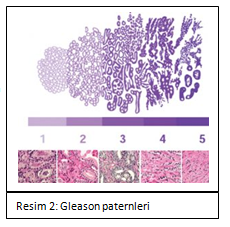
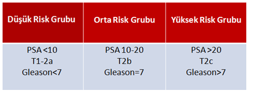
In localized (limited to prostate) prostate cancer, radical prostatectomy surgery, radiotherapy or radiotherapy + androgen deprivation therapy for a limited time is decided by talking to the patient according to the risk groups.

Due to the slow course of cancer, “delaying treatment” called “active surveillance” is another alternative. For this aproach, patient selection and compliance with follow-up is extremely important.

Focal therapies in localized cancer can also be applied, although the level of recommendation is low.

In locally advanced (overflowing from the prostate but not spreading to other organs) prostate cancer, radical prostatectomy surgery or radiotherapy + 2-3 years of LHRH analogue treatment is preferred.

Orchiectomy (removal of both testicles) or LHRH analogue combination is used in metastatic disease.

In metastatic disease, it has now been shown that orchiectomy or LHRH analogues with chemotherapy prolongs the life expectancy by an average of 13 months. Local treatment with radiotherapy or surgery in low-volume metastases and radiotherapy for metastases have also been among the applications of treatments in recent years.

Why are the testicles of the patient removed in metastatic cancer?

Prostate cancer is a “testosterone” dependent cancer. Testicles, the main source of testosterone in the body, are removed to slow cancer. With this treatment, the cancer becomes silent for approximately 24-36 months. However, cancer cells find molecules other than testosterone to reproduce at the end of this period and the disease is defined as “castration resistant” after this stage.

As with orchiectomy, very low testosterone levels can be achieved with LHRH analog/antagonist drugs without removing the testicles. These drugs inhibit the production of testosterone from testicular tissue.

Does chemotherapy have an effect in the treatment of metastatic prostate cancer?

Chemotherapy in prostate cancer is the first choice in castration-resistant metastatic disease.

In 2015, CHARTED and STAMPEDE studies showed that there was an approximately 13-month prolongation in life expectancy with chemotherapy in the presence of a newly diagnosed hormone-sensitive but high-volume matastatic disease (visceral organ metastasis, 4 bone metastasis or extra-vertebral bone metastasis). Thereupon, the treatment algorithm in prostate cancer has changed and subsequent studies have shown that some androgen receptor blocker drugs (abiraterone, enzalutamis ..) increase the life span just like the chemotherapy (Docetaxel). Today, chemotherapy / immunotherapy is used in combination with “androgen deprivation therapy-ADT” (LHRH analogue / antagonist) in metastatic prostate cancer, regardless of low or high volume.

Does radiotherapy have an effect in the treatment of metastatic prostate cancer?

The results of the STAMPEDE study, published in 2018, showed that in patients with low-volume metastases (<4 bone metastases and no extravertebral metastases, no organ metastasis), radiotherapy to the prostate prolongs the life span. In these patients, radiotherapy for prostate and/or metastasis can be applied.

Does surgery have an effect in the treatment of metastatic prostate cancer?

There are studies showing the effectiveness of local treatments (Radical prostatectomy or radiotherapy) in metastatic prostate cancer.

Patients’ reservations about treatments

            Incontinence

After radical prostatectomy, urinary incontinence is one of the most anxious issues for patients. The early urinary incontinence after surgery decreases over time with the strengthening of the auxiliary muscles. In the first year after surgery, the rate of complete dryness of patients is reported between 80% and 97% in different clinics. The age of the patient, the presence of urinary retention problems before the operation, the protection of the bladder neck during the procedure and the experience of the surgeon are important factors affecting postoperative urinary incontinence.

            Erectile dysfunction

Another situation that can be seen after radical prostatectomy and causes anxiety in patients is the problem of erection.  Erection problems at different levels can be seen in 70-75% of patients after surgery. Of course, the surgical technique (nerve-sparing approach) and the preoperative erection capacity of the patient significantly affect the postoperative erectile dysfunction rate.

Also after radiotherapy, nearly half of the patients can have erection problems.

After LHRH agonists or orchiectomy surgeries, the testosterone level decreases too much, resulting in erection problems and reducing libido.